877/1, Deewans Road Lakshmipuram,Mysore 570004 Karnataka,India

-
9845971425
contact to
-
ukehkvr@gmail.com
Email through

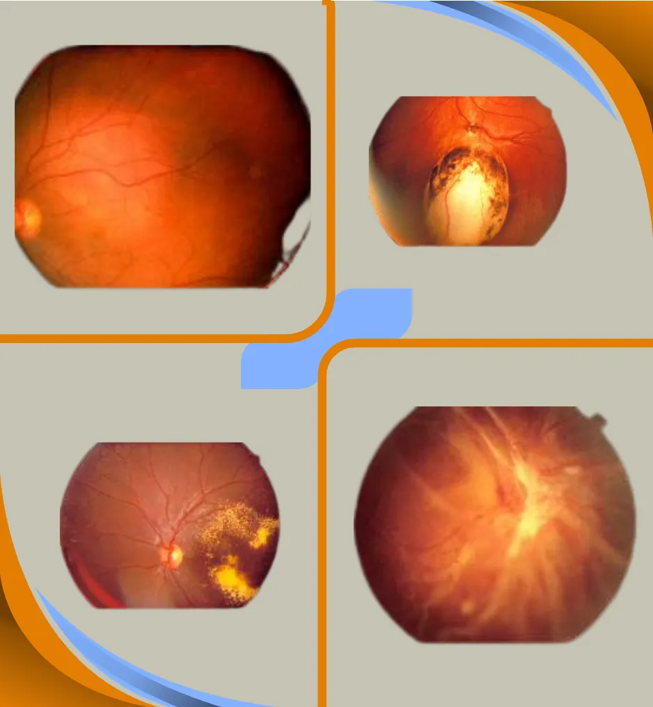
Viteroretinal Surgery
At Usha Kiran Eye Hospital, our Vitreo-Retinal Services provide comprehensive medical and surgical management for a wide range of vitreo-retinal diseases. We specialize in treating sight-threatening conditions such as diabetic retinopathy, retinal vascular occlusions, age-related macular degeneration (ARMD), penetrating ocular injuries, retinal detachment, and rare genetic disorders. Our services integrate cutting-edge diagnostic technology and advanced therapeutic interventions to ensure the best possible outcomes for our patients.
Disorders Treated
Diabetic Retinopathy:
Diabetic retinopathy is a complication of diabetes and a leading cause of blindness. It occurs when diabetes damages the tiny blood vessels inside the retina which comprises the light-sensitive tissue at the back of the eye. A healthy retina is necessary for good vision. If you have diabetic retinopathy, at first you may notice no changes to your vision. But over time, diabetic retinopathy can worsen and cause vision loss. Timely intervention in the form of laser , intraocular injections or apt surgical treatment will arrest the progression of the disease, along with adequate diabetic systemic control and close follow up.
Age Related Macular Degeneration:
In macular degeneration, the light-sensing cells of the macula malfunction and may over time cease to work. Macular degeneration occurs most often in people over 60 years old, in which case it is called Age Related Macular Degeneration (ARMD). ARMD can be “dry” or “wet” depending on the degree of damage. While dry ARMD can be observed – the wet type will need timely intraocular injections aimed at halting the disease progression.
Flashes and Floaters:
Light flashes are sometimes caused by mechanical stimulation of the retina. A variety of conditions can cause it, including posterior vitreous separation and retinal tears. People who have a high minus refractive error or myopia are most prone to developing retinal thinning holes and tears. A regular follow up with the retinal specialist will help in picking up the disease in its early stages.
Floaters are black coloured small objects that are seen in the field of view of the patient’s eyes.Sudden onset of these floaters can sometimes indicate retinal tear / bleeding into the cavity of the eye and has to be consulted with the retina specialist on an emergency basis.
Retinal Detachment:
Retinal detachments often develop in eyes with retinas weakened by a hole or tear. This allows fluid to seep underneath the retina , weakening the attachment so that the retina becomes detached like wallpaper peeling off a damp wall. When detached, the retina cannot form a clear picture and the.
vision becomes blurred and dim. The retinal tissue starts to lose its function very quickly . Hence these symptoms should be brought to the notice of the retina specialist at the earliest so that a timely laser or surgical intervention can restore the vision to the best possible extent.
Retinal Arterial Blocks:
A retinal artery occlusion occurs when the central retinal artery or one of its branches gets blocked. This blockage is typically caused by a tiny embolus (blood/fat clot) in the bloodstream. The occlusion decreases the oxygen supply to the area of the retina nourished by the affected artery,causing permanent vision loss. Patients usually have a sudden loss of vision – typically when they wake up from sleep. Retinal artery obstruction is comparable to a stroke in the eye. The damage can be quite severe, depending on the extent to which the blood flow has been disrupted.People with co-morbidities like diabetes, hypertension , underlying blood disorders are all prone to developing this condition..
Retinal Venous Blocks:
Central and branch retinal venous occlusions occur when there is a congestion to blood flow and an increase in backpressure on the central retinal vein. It causes a variable degree of visual loss and can be easily diagnosed by a retinal examination. It is commonly seen in hypertension and diabetes. Sometimes it can be seen in people with blood clotting disorders. An early OCT scan of the retina can help the retina specialist pick up a macular edema due to the occlusion and can be treated with intraocular injections aimed to salvage the vision to the best possible extent.
Other Disorders Treated are
- Vitreous hemorrhage
- Cystoid macular edema
- Macular hole
- Retinitis pigmentosa
- Posterior uveitis
- Ocular trauma
- Intraocular tumours
- Central serous retinopathy
- Retinoblastoma
- Hereditary retinal degenerations
- Others
Procedures & Advanced Diagnostic Tools
Our Vitreo-Retinal Department utilizes state-of-the-art equipment and techniques for diagnosis and treatment
Outpatient Procedures:
- Indirect Ophthalmoscopy- To have a detailed binocular 3-dimensional view of the retina. The patient is asked to lie down and with an indirect ophthalmoscope, we look at the full extent of the retina. Sometimes, it may be needed to press on the lids to get a good look at the edge of the retina.
- Stereo biomicroscopy- This is done for a magnified examination of the central retina and optic nerve.
- Digital fundus camera and imaging-To take serial pictures to evaluate any subtle changes that may occur with time. A key for all diseases in which we may require observation and follow up.
- Fundus Fluorescein angiography(FFA) and indocyanine green angiography(ICGA)-A dye is injected into the veins of the hand and serial pictures are taken with the Heidelberg ™ imaging apparatus. This investigation set helps us obtain information regarding retinal andchoroidal blood circulation which is a gold standard for diagnosis of many retinal and choroidal disease states.
- Optical coherence tomography(OCT)-It is one of the most important diagnostic tool to evaluate the depth and extent of almost nearly every retinal condition. It gives us a detailed evaluation of all layers of the retina and choroid similar to a tissue study under the microscope but in a non invasive manner. It is based on reflectance of light waves of a certain wavelength from the retino-choroidal tissues.
- Optical coherence tomography angiography( OCTA)-This investigation helps in understanding the blood circulation of the retina in a non-invasive manner and can help us avoid the FFA/ICGA in certain specific conditions -thus eliminating the need for intravenous dye injection.
Treatment Techniques:
- Ultrasonography-Standard of care for vitreo-retinal disease diagnosis in hazy media like cataract and vitreous hemorrhage. It is a useful aid in cases of uveitis.
- Green laser photocoagulation-This is used for treatment of diabetic retinopathy, peripheral retinal degeneration like lattice degeneration, retinal breaks, etc
- indirect Ophthalmoscopy-Is another type of laser delivery system for treatment of peripheral retina, retinopathy of prematurity (ROP), recent post operative cases etc.
Why Consider Vitreoretinal Surgery?
Vitreoretinal surgery offers vital solutions for patients facing serious retinal conditions. Our advanced microsurgical techniques, such as vitrectomy and laser photocoagulation, help restore vision and prevent further visual decline. From reattaching the retina to repairing macular holes and addressing complications of diabetic retinopathy, our team is dedicated to preserving your vision and enhancing your quality of life. By integrating world-class expertise, innovative technology, and compassionate care, Usha Kiran Eye Hospital remains a leader in the field of vitreo-retinal treatment.
Vitreoretinal Surgery Benefits
Vitreoretinal surgery offers numerous benefits for patients with serious retinal conditions. Here are the key advantages:
- Improved Vision Stability and Clarity
- Prevention of Further Vision Loss
- Enhanced Quality of Life for Patients with Retinal Disorders
- Effective for a Range of Complex Retinal Conditions
- Minimal Pain and Discomfort During Recovery
- High Success Rates with Advanced Techniques
- Preservation of Visual Function for Daily Activities
- Potential for Long-Term Vision Improvement
Our Video Gallery
Explore our collection of informative and engaging videos that showcase our services, patient testimonials, and educational content about eye care. Discover how Usha Kiran Eye Hospital is dedicated to providing exceptional eye care and enhancing the vision of our patients.